Product Description
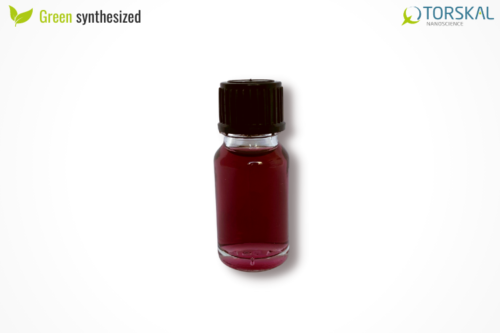
Gold nanoflowers are synthesized following a process patented by TORSKAL using crude and/or purified extracts from plants. This green, reproducible synthesis leads to very stable gold nanoflowers without hazardous chemicals. Plant extracts have a role in both synthesis and stability of the gold nanoflowers. Thoroughly washed nanoflowers are suspended in water or can be dispersed in other solvents or buffers.
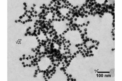
Each batch of gold nanoflowers are extremely characterized using techniques including transmission electron microscopy (TEM), dynamic light scattering (DLS), zeta potential, and UV-Visible spectroscopy.
Applications of our gold nanoflowers
Product Specification
| Parameters | OD : 1 | OD : 10 | OD : 50 |
|---|---|---|---|
| Diameter (TEM) (nm) | 49.57 | Same | Same |
| Size disparity (+/- nm) | 9.41 | Same | Same |
| Diameter deviation | 15% | Same | Same |
| SPR Peak | 550 – 555 | Same | Same |
| Particle concentration (NP/ml) | 3.4E + 12 | 3.4E + 13 | 1.7E + 14 |
| Mass concentration (mg/ml) | 4.13E – 2 | 4.19E – 1 | 2.10E + 00 |
| Particle molar concentration | 5.65E – 11 | 5.65E – 10 | 2.825E – 09 |
| Zeta potential (mV) | -33.8 | Same | Same |
| Zeta potential deviation | 10% | Same | Same |
| Particle volume (nm3) | 6.37E + 04 | Same | Same |
| Particle surface (nm2) | 7.72E + 03 | Same | Same |
| Surface/Volume ratio | 0.121 | Same | Same |
| Solvent | DIH = 18MEG DI Water | Same | Same |
| Stability | 3 months | Same | Same |
| Storage temperature | 4 – 8 °C | Same | Same |
| Color | Purple | Same | Same |
Physico-chemical Characterization
")
Extinction spectrum of the solution of FAuNPs 50
")
Size distribution of FAuNPs 50 determined by transmission electron microscopy image